Tuesday 28 October 2014

Ancient Proboscideans – Extinct Elephants and their kindred.


Proboscidea is the order of mammals with trunks, and their more primitive relatives with “proto-trunks”. Today the only living members of this order are in the family Elephantidae, the living Asian, African savannah and forest elephants. These are magnificent megafauna in themselves, treasures of any zoo which has them, butchered for their incisors and occasionally used in human warfare. But there is much more to Proboscidea than the living elephants as this order has a total of around 164 mostly extinct species, which diversified into all sorts of bizarre forms, such as the four tusked deinotheres and the shovel-tusked Platybelodon. These fantastical, mighty beasts are only known to us through the fossil record. It is tempting to compare the largest Proboscideans, to the herbivorous dinosaurs, especially the sauropods, as they fill the niche for large browsing animal. I have no chance of discussing all these species here, but I will mention some of the most outlandish and interesting Proboscideans less familiar to us than elephants and mammoths.

Animals with 3m long teeth don't drop out of the sky, they as an order have a history stretching back into the Paleocene, starting with an apparently humble creature the size of a rabbit. The evolutionary process basically involves a gradual increase in body size, growth of tusks and size of head. The heavy tusked head could not be supported by a long neck, and as their legs were long they could not reach the ground with their mouth to eat and drink. This problem was “solved” by the elongation of the muscular nasal cavity, forming a trunk. These processes were not so smooth and continuous, and the journey had many turns from this path.


A reconstruction of Eritherium.

One of the earliest Proboscidean is Eritherium which lived 60 million years ago, the remains of this creature was first found in phosphate deposits in Morocco in 2012. The Eritherium remains show no sign of a trunk, which is formed by the evolutionary fusion of upper lip and nostril, which would be evident in an enlarged naval cavity. It is a Proboscidean without a proboscis, but probably had a very mobile upper lip, a little like the unrelated tapirs. So what makes this animal a Proboscidean? A number of features which were elaborated in later Proboscideans are present in Eritherium, including enlarged incisors, which would eventually sprout forth to create tusks, and simple lophodont molars (molars with ridges perpendicular to the jaw line). It was 5kg in weight, but this outweighs most other Paleocene mammals, many of which were decided shrew-like. Eritherium was probably somewhat aquatic, like many of the Proboscideans including the swamp dwelling American Mastodon and modern elephants who use their trunks as “snorkels” in order to swim up to 48km offshore. It is likely that the common ancestor of Proboscideans, Sirenians (sea cows) and the extinct Desmostylia were fully aquatic.


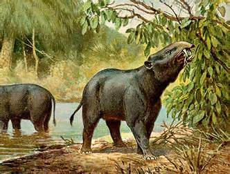
A reconstruction of Phiomia

A later group of Proboscideans is Phiomia, a direct relative of modern elephants, which lived between 35 and 25 million years ago. This was a larger animal, 2m high at the shoulder. Remains show evidence of a short rudimentary trunk (based on the larger nasal cavity) and enlarged incisors on the lower and upper jaws, meaning it had two pairs of tusks. Tusks are simply enlarged incisors, about a third of it's length, the pulp cavity, is embedded in the skull. The visible tusk is the ivory made of dentine covered by enamel. After it has shed it's milk tusks a Proboscidean maintains and grows it's tusks throughout it's life. In observed species the male has the longest tusks, as befitting their primary use in fighting, but they are and were also used to strip bark, defend from predators and in species like the woolly mammoth they may have been used to break up ice to find food.


A skull of Deinotherium at the Natural History Museum

The branches of evolutionary tree which did not lead to elephants contain more baroque Proboscideans, including two groups which lived at approximately the same time, Deinotherium and Platybelodon. The skull of Deinotherium is a disturbing thing to bump into in a museum, no doubt horrifying for an ancient person, with no ideas about deep time, to find in the wild. The skull appears to have two horns curved backwards out of it's chin, like a demonic beard presumably used for gouging. If an ancient person who expects all animals to have forward facing eyes like they do, sees the enlarged nasal cavity at the front of the skull, it could be mistaken for the skull of a monster. Indeed, the ancient myths of the Cyclops could be inspired by the discovery of Proboscidean fossils in Greece. Deinotherium itself lost it's upper pair of tusks and maintained the lower tusks (the opposite of modern elephants). As you can tell from a reconstruction of these creatures, the lower tusks hid quite discretely under the trunk which filled the nasal cavity at the front of the animals head, making it look less horrifying. They were quite primitive, lacking the sequential teeth eruption of later Proboscideans.

The skull of Deinotherium is a disturbing thing to bump into in a museum and no doubt horrifying for an ancient person, with no ideas about deep time, to find in the wild. The skull appears to have two horns curved backwards from it chin, like a monstrous beard, used for gouging. Indeed, the ancient myths of the Cyclops could be inspired by the discovery of Proboscidean fossils in Greece, due to the fused external naris resembling an eye socket. Even the name Deinotherium is from the Greek for “terrible beast”. In reality, Deinotherium lost it's upper tusks and maintained it's lower tusks (the opposite of modern elephants), and probably used these curved tusks to scrap bark from tree. The lower tusks would have hid quite discretely under the trunk which extended from the external naris, making it look less horrifying.


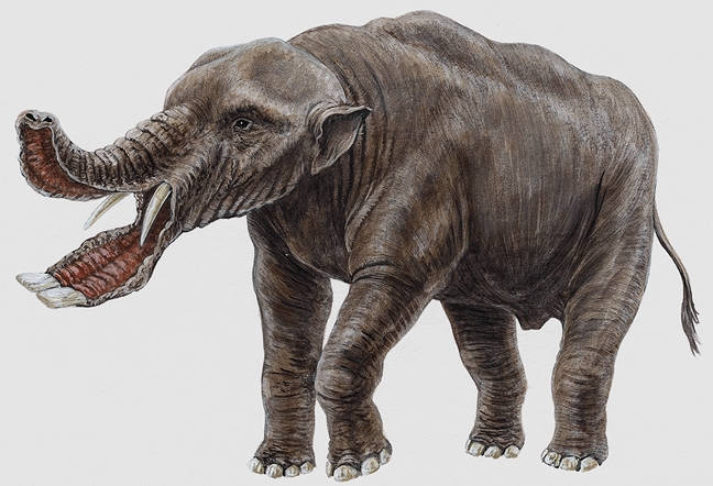
A reconstruction of Platybelodon

Platybelodon is in the same family as Gomphotherium and is not a direct ancestor of elephants. It lived about 20 – 8 million years ago and retained all four tusks. The lower tusks flattened out so the each tusk met and formed a sort of “shovel” shape with a deep scoop at the end, which only developed in adulthood. There is speculation about the use of it's tusks. It was originally thought that it they used their “shovel” to scoop through the mud to collect plants; a semi-aquatic lifestyle familiar to Proboscideans. The two lower tusks end with a V-shaped sharpened tip, analysis of the pattern of wear suggests they were used in a scythe-like manner to cut down branches and to strip bark from trees. Palaeontologists removed them from their presumed habitat of lake-side bogs and place them in a more arboreal habitat. The shovel tusks were an adaptation to a crowding niche, as with several genera of Proboscidean in this area at the time, Platybeldon had to specialise to survive.


A reconstruction of Gomphotherium

Another key part of the future elephant physiology was put in place in another of the elephants' direct ancestors, Gomphotherium, living from 20 to 15 million years ago. They retained the four tusks, with the lower tusks being slightly flattened, and show a basic form of sequential tooth development. This is when, due to the stresses caused by eating grasses containing silicon particles, a tooth erupts from behind the existing teeth to replace the one which had eroded at the front. The erupted tooth would be larger than the previous ones, therefore allowing the jaw to continue to grow throughout the animals life. Gomphotherium probably had three teeth in each side of it's jaw, but later species had only one tooth in each side of the jaw. Gomphotherium was a member of the well travelled family Gomphothere, which includes the genera Cuvieronius and Stegomastodon, some species of which travelled as far as South America.


A sketch of American Mastodon Molars, my work

Mastodons were, despite their common confusion with mammoths, very different animals, Mastodons diverged from the line that would lead to elephants after Phiomia, separating them from mammoths by about 20 million years of evolution. The earliest Mastodons were the Losodokodon which lived from 27 to 24 million years ago in East Africa, later radiating throughout Europe, Asia and North America. The most famous Mastodon species is the American Mastodon, whose genus Mammut arrived in North America 11 million years ago. The American Mastodon was around 2.7 metres at the shoulder, small compared to the neighbouring Columbian Mammoth. It was quite stocky with a deep chest and probably quite muscular. It's tusks were up to 2.5 metres long in adult males and curved upwards and slightly outwards, less elaborate than tusks of mammoth. The name Mastodon means “breast tooth”, showing the lumpy nature of their molars. Elephant and mammoth molars are relatively flat and ridged, whereas mastodon molars have rounded cusps, which caused early naturalists to speculate that they were terrifying beasts who caught prey with their tusks. The reason each of these groups have significantly different teeth is because they were used to eat different foods, none of these animal foods. Mastodon teeth were used to crush leaves and twigs in forests, whereas mammoths grazed grassland.


My drawing of a cave painting of a woolly mammoth

The genus believed to be the ancestors of modern elephants and mammoths is Primelephas. These creatures had four tusks, though these were smaller than in Gomphotherium. This is because Primelephus did not need them to shovel though the mud, it had moved to a grassland habitat similar to modern elephants on the savannah or mammoths on the steppe. This genus split into three genera, Loxodonta (African elephants), Elephus (Asian elephants) and Mammuthus (mammoths). Mammuthus belongs to the Elephantidae family, making it as much of an elephant as Loxodonta and Elephus.


Bibliography


Understanding proboscidean evolution: a formidable task- Jeheskel Shoshani www.sciencedirect.com/science/article/pii/S0169534798014918

I found the exhibition Mammoths: Ice Age Giants at the Natural History Museum very informative, as well as the book of the same name by Adrian Lister.


This was an entry to Rockwatch's Young Writer 2014 competition. 


Tuesday 23 September 2014

Rabies: the 11,000 base pairs that can ruin the brain


Lyssavirus

On the 16th October 2004, a teenage girl was brought to the Children's Hospital of Wisconsin and tested positive for a disease that all the hospital staff knew had a 100% mortality throughout all recorded history. Her parents were given two options, to put her in a dark room in the Hospital to die or take her home to die.
That September, the girl, Jeanna Giese, was bitten by a strangely acting bat on her left index finger. On 13th October, Jeanna felt ill and had tingling in her left arm, but from there deteriorated rapidly. She couldn't walk, her left arm twitched uncontrollably and her speech was slurred. Hours after arriving at the Hospital she became stuporous. She had rabies, and appeared to be going the same way as all other rabies victims.

Rabies is caused by viruses of the genus Lyssavirus, all of which as around 11,00 base pairs long. The disease is zoonotic, meaning it's host is non-human animals. Rabies can infect almost all warm-blooded animals, though the disease is mainly transmitted to humans via contact with the saliva of bats, dogs, raccoons and some other animals. Rabies is a peculiar virus. The classic virus enters the blood stream and circulates until it reaches it's target where it replicates. Rabies does not take this blood route; instead it takes the nervous route. The virus binds to the nerve nearest the bite and inches it's way up the periphery nerves and into the central nervous system until it gets to the brain. It travels under 2 cm a day, therefore it will take longer to travel to your brain if you are bitten on the toe, maybe months, but when bitten on the face it could take a week. This window of time is crucial, as this is when you can administer a vaccine to cause the body to mount an immune response which destroys the virus. When the virus reaches the brain the vaccine is useless, as the virus will now act at a swifter speed that any normal immune response can.
What the virus does in the brain is unknown. The two main theories are that it either over-excites the brain so that it cannot carry out normal functions and the person dies (known as excitotoxicity) or that it simply inflames and kills neural tissues.
This is what is does to a patient's biology, but it does much more to the patient's behaviour.
30% of patients experience paralytic rabies, where they are paralysed and then slip into a coma and die of heart or other organ failure.
The rest have a different fate. A few days before death they experience convulsions, hallucinations and explosive rage. The brain cannot regulate their body; throat spasms cause wild cries and they can die by drowning in their own blood or organ failure. This behaviour is very animalistic and uncontrollable, it could explain the werewolf myth.
But the truth is more chilling. A person's behaviour is not driven by human logic but by a virus' need to replicate; a sentient being acts this way because of a simple virus. Patients experience hydrophobia so they can't drink, which then dilutes their saliva and they cannot swallow, meaning that their virus-rich saliva can only go one way. They are aggressive so they might bite and spread the virus. This is an assault to our idea of free will; if a micro-organism that isn't really alive can change our behaviour so much, then are we as autonomous as we like to think? It is one of the most undignified ways to die imaginable.
Therefore, Jeanna's doctor, Rodney Willoughby, was not content to see his patient die without doing something. He thought that, if rabies was an excitotoxic disease, then if the immune system was given enough time to catch up with the virus so antibodies were produced in time so the patient could recover. So Dr Willoughby put Jeanna into a coma and waited. This was an enormous risk, as she could be brain-damaged if she even survived at all. 7 days later she was brought out of a coma and though weak and frail, alive, against the entirety of scientific consensus. 
She had produced enough antibodies in enough time for her immune system to fight off the virus, the first person known to have encountered rabies and come out alive.
Jeanna was very weak, and had to be treated with drugs including ketamine and antivirals. She has been through many years of rehabilitation and treatment, but today is healthy and spends her time with her sled dog team, and despite her experiences still has a love for bats (as we all should). 
There is, however, controversy as to the effectiveness of Dr Willoughby's treatment, which is called the Milwaukee protocol and has been used on a handful of other patients, with only 4 of 35 surviving, which is still an impressive survival rate for rabies. It has been suggested that Jeanna and the others survived because they were genetically "fitter" to withstand rabies, they were infected with a weakened form or rabies or bitten on a "safer" area or all of these together. There are even reports of some populations (mostly remote, Amazonian populations) found with rabies anti-bodies in their blood (with no signs of rabies, and they probably couldn't have recovered without the Milwaukee protocol), indicating a genetic resistance to rabies, in a similar way that some tropical populations have a genetic resistance to malaria. 

Bibliography


http://www.ncbi.nlm.nih.gov/pmc/articles/PMC3414553/ : This is by Rodney Willoughby himself. 


Friday 29 August 2014

Notes on Objects: Longhorn Beetle and Ammonite

I volunteer as an object-handler at Manchester Museum (I am there on Wednesdays from 11am to 1 pm, at the table near Nature's Library), which involves inviting people to handle certain objects from the museum's archives and talking to them about it. I often have a taxidermied fox, which is a good hook to bring people in, and they often have the slightly philosophical question, "Is it real?". It is real fox fur (and maybe claws?) but the insides and eyes aren't real, the insides are a polyurethane mold and the eyes are plastic. So it is a semi-real fox. Many people are very surprised that it feels so soft around the ears, a bit like a dog.
But my two favourite objects are the Giant African Longhorn Beetle and an unidentified ammonite, and I will give some further information about them here.


Giant African Longhorn Beetle (Petrognatha gigas)


This is a insect of the family Cerambycidae (the long-horn beetles), and the "horns" of it's name are it's antennae, which are used in insects for sensory purposes, including orientation, detecting sound and sensing chemicals. The Giant African Longhorn Beetle lives in dead acacia trees, and it is well suited to this, as it's long antennae and limbs look like the twigs of the acacia tree. The spikes on it's thorax (the "neck") and top of the abdomen look very like the prickles of the acacia tree.


Beetles are insects with “sheathed wings” according to the literal translation of their scientific name ColeopteraAll insects have two pairs of wings, the hardened forewing (elytra) and the hindwing, which is a fairly standard insect wing. The elytra protects the hindwing in flying species. In ground beetles, the elytra are fused together, therefore making them flightless and confined to the ground. All beetles have a hardened exoskeleton, which is made of sclerite plates separated by a rigid joint (suture) which provides both armour and flexibility. Not a lot is known about Giant African Longhorn Beetles as they have not been studied much, though they do have a particular way of breathing known as Bernoulli suction ventilation, see this article to learn more. 


  Cerambycidae is a large family of over 20,000 species. The largest member of this family is the Titan beetle (Titanus giganteus), which has a maximum body length of 16.7 cm. The longest beetle of all is the Hercules beetle (Dynastes hercules) which is up to 17.5 cm long and can lift up to 8kg over 850 times their own weight. Hercules beetles are rhinoceros beetles, so named because the male has two large horns on it's head, adding to it's length, which are used for fighting.  The larvae of this family are called roundhead borers, as they bore into living or recently felled wood, and are considered a pest by the logging industry, though the larva of the cactus longhorn beetle bore into prickly pears and chollas.
There are around 400,000 known species of beetles (25% of all know animal species), and there are probably over 1 million total known and unknown beetles in the world. 


Ammonite (of unknown classification)


 Ammonites were shelled, marine cephalopods (the same group which octopuses and squid belong to) which existed from the mid Devonian (400 million years ago) to the late Cretaceous (66 million years ago), in the KT (Cretaceous-Tertiary or Cretaceous- Paleocene) Mass Extinction which was probably caused by the effects of an asteroid impact and also caused the extinct of the non-avian dinosaurs. The ammonite I have on my handling table  is 160 million years old, placing it in the Late Jurassic (in the Oxfordian age), which is 95 million years older than T. rex. Ammonites as a group are much older than the oldest dinosaur (probably Eoraptor, which is 231.4 million years old), and even older than four-legged animals (Tetrapods) which evolved after 395 million years ago.  The white stuff in the sections of the shell is cacite crystals, which were formed when the shell was being fossilised. Water with dissolved salts in it got into some of the outermost sections of the shell, and as the shell was being compressed under the weight of sediment on the ocean floor, the water evapourated and left behind the salts, which grew into crystals. 

They are named ammonites due to their resemblance to the ram-like horns of the Egyptian God Ammon or Amun.


All regular ammonite shells grew out from the centre (the umbilicus) in a spiral in one plane, becoming gradually thicker as it grew. It grew by adding chambers on the shell from the umbilicus, and when it “moved” from an old chamber to a new one (the living chamber), it sealed off the old chamber except for the siphuncle, a “tube” which extended through the shell. The siphuncle could be used to change the air pressure in the sealed-off chambers (known as the phragomocone), so it could rise or descend, rather like a submarine does. The earliest phragomocene chambers tend to get less sediment in them, therefore it is common for crystals to grow in these sections. My ammonite has calcite crystals (which are a very common crystal) in a lot of the phragomocene chambers.


One of the largest ammonites found, Parauzosia seppenradensis


Ammonites would probably have had ten arms (all cephalpods have arms, not legs). It's arms may have surrounded hard aptychi, which may have been the jaw apparatus of the ammonite, similar to the beak in other cephalpods, or the aptychi could have been a sort of head shield, which is seen in living nautiloids. There is very little fossil evidence of the soft parts of ammonites, though it is likely that, in addition to the ten arms, they had an ink sack and a hyonome (see below).

Ammonites are fairly distant relatives of the Nautiloids (which include the living Nautilus). The Ancestors of Ammonites branched off from the Nautiloid line (specifically straight-shelled Bactritid nautiloids) before 400 million years ago.

What is the difference between the Nautiloids and the Ammonites, then?
The basic difference is the difference in the sutures. These are the lines where the walls which separate each chamber (called the septa) meet the wall of the shell, which can be seen by looking at the outside of the shell. Nautiloid sutures are generally gentle curves, whereas Ammonite sutures are more wavy. The suture lines are determined by the shape of the septa when it meets the wall of the shell. The difference in the suture lines are caused by the different shapes of septa in Ammonites and Nautiloids. Ammonite septa are more wavy and to a varying extent convex from the front of the shell, whereas Nautiloid septa are smoother and more concave from the front. The shape of the septa can be observed when the ammonite shell is in cross-section. The wavy suture lines enabled ammonites to withstand high pressures whilst having a thinner shell than Nautiloids, though both subclasses were able to living at depth (Nautiloids at a deeper depth than Ammonites).

Ammonites and Nautiloids both have a siphuncle, but the siphuncle of Nautiloids is in the middle of the septa, whereas the ammonite siphuncle is on the outer edge of the shell. 


Saturday 9 August 2014

The evolution of the oocyte


A human egg cell, the corona radiata is on the outer surface, the zona pellucida is the clear ring behind it. 


Oocyte is the name for an immature egg cell (ova, in animals), an oocyte only becomes an egg cell finally when it is fertilized. 
The oocyte and the spermatozoa (sperm cell) are the sex cells of organisms which reproduce sexually, that is by combining the genetic material of two different organisms. Yet, the very earliest cellular organisms must have reproduced asexually, so why does the vast majority of complex organisms produce sexually exclusively? What caused this change, and why did it proliferate?

There is much speculation on why so many organisms switched from asexual to sexual reproduction. It is likely that sexual reproduction arose in an ancestor of the eukaryotes (organisms with more complex cells), deep in the Precambrian. But sexual reproduction has an immediate flaw, only half the population are capable of producing offspring physically, the females. An asexual population would quickly out-compete a sexual one purely by virtue of being able to grow at twice the rate of a sexual one. This is known as the “two-fold cost of sex”. However, this overlooks the role of the male. Males provide genetic diversity to a particular population, as when the sperm and ovum haploid genomes recombine in fertilization to form a zygote with a diploid genome, and therefore two copies of the same gene from each parent. In recombination, genetic “mistakes” in the sequence of base pairs should be corrected. Hybrids between two populations of the same species tend to be fitter genetically, a process known as heterosis (this is not always the case in all hybridizations, however). Therefore, an organism produced sexually may be more resistant to disease than asexually produced organisms. This is probably why almost all complex organisms (except very few, like the New Mexico whiptail lizard, which can also be created by hybridizing two other whiptail species) reproduce sexual, as they need to compete in a more dynamic ecology, so need the fast acquisition of advantageous traits in the whole population that only sexual reproduction. 


Isogamy and Anisogamy 


Some of the more primitive single-celled organisms reproduced by fission, which is regular cell division into genetically identical offspring cells. However, most single celled organisms swap genes, occasionally, through genetic recombination or the transfer of a nucleus mostly with organisms from a separate population, which has accumulated different genetic mutations to it's population. This is probably how sexual reproduction arose in the earliest organisms. Eventually specific cells began to specialize, by carrying the haploid genome of the organism, so it would combine with the haploid genome of another organism to create a diploid organism. This is this basis of sexual reproduction.
The gametes in these early sexual organisms would have appeared the same, only the haploid genome would be different between the two. This is known as isogamy, where each gamete has the same morphology. The gametes in some organisms began to specialize further, into anisogamy, where one gamete is larger than the other. One modern partially isogamous species is the green algae Volvox. In a study, colonies where all the gametes were small (isogamy) produced a smaller population, whereas anisogamous populations were larger. This shows that when there is one large gamete, which has a larger energy reserves (yolk granules in the cytoplasm) but is slower, and one small gamete, which can move faster but does not have large energy reserves, population sizes are larger. Anisogamy means that one gamete, the oocyte, can specialize in providing a larger energy supply for the zygote after fertilization and the other, the spermatozoa can specialize in being fast and numerous. This is a particular type of anisogamy known as oogamy. 
Due to the large tax on energy resources for the female there is a great difference between ovum and sperm number, there are around 400 ovum ovulated by a woman who has reached menopause compared to 200-500 million spermatozoa per ejaculate for human males, and up to 8 billion per million per ejaculate for domesticated pigs (who have been breed to be as fertile as possible).


Egg Laying 


The first vertebrates to have ova were reptiles, as when on land they could not reproduce by spawning. In reptiles, and later in birds, the ova once fertilized forms protective layers (the egg shell) and is passed out through the oviduct. In some of these species, the embryo is then kept warm (incubated) by mostly the mother (the father and mother Whooping Crane alternatively incubate the egg). This is known as ovipartity, and the embryo receives nutrition only from the nutritive yolk, meaning than in oviparous animals, more energy is needed to create the ovum than in viviparous animals (who have live young). But after fertilisation, there is less need for parental care, therefore enabling more offspring to be produced. The offspring, due to lack of long-duration parental care, have a higher mortality rate, but more are produced in relation to the amount of energy the parents lose in reproduction. Only a small number of mammalian species produce eggs (the monotremes) including the duck-billed platypus, which are descended from the some of the earliest mammals and hence having features common and almost unique to both mammals and reptiles.


The Zona Pellucida


The oocyte has multiple barrier to stop polyspermy (multiple sperm entering the oocyte, therefore producing a cytoplast with around double the normal number of chromosomes for the species, which would be unable to survive). Polyspermy may arise from having sperm that is too efficient at surviving and fertilising the oocyte, therefore a male with very efficient sperm may not pass on it's genes due to his offspring's inability to survive.
Sperm that is more efficient at fertilising an oocyte does not mean that it is beneficial to the female (for reasons as seen above) as this sperm may not have the best genetic material to pass onto successive generations. If mammals and reptiles evolved using spawning as a reproductive method, there would be all sorts of genetic problems as the only “screening process” would be that the sperm would be from an animal that had reached puberty. This gives rise to a very complex genital system in polygamous species, such as that seen in the Pekin Duck. The female Pekin Duck has a maze-like vagina to be able to “pick” the best sperm for reproduction. To counter this the male has a “corkscrew”-like penis and only the males with the precisely shaped penis for that one female's vagina will fertilise the oocyte. 
The first barrier to the sperm is the zona pellucida, then the plasma membrane, which also protect the fertilized ova on it's journey through the oviduct (or in the water in fish and amphibians). The zona pellucida is an essential feature, if it is not present the female will be infertile. It stops sperm from other, genetically incompatible species from binding and also emits chemicals that guide the sperm cells towards the oocyte. In ovipartial species, when the nucleus of the sperm and oocyte fuse, the zona pellucida signals for a release of calcium ions which eventually transform into the egg shell. For fertilization to occur the nucleus of the sperm needs to bind with the zona pellcida and “bore through” it, fusing in the oocyte's cytoplasm. After fertilisation the cortical reaction is triggered, which causes the plasma membrane to fuse and become impermeable to sperm. Without the zona pellucida, gametes from incompatible species can fuse, though the offspring are not viable.
Genes encoding on the surface of gametes are some of the most rapidly evolving genes, even faster than those responsible for immune responses, which have to adapt quickly due to microbe attacks. If the oocyte is "under pressure" to reduce polyspermy, the proteins on the surface of the oocyte will change, so that fewer sperm are compatible with the oocyte, meaning there is a smaller probability of polyspermy.


Bibliography


Thursday 7 August 2014

Mammoths: Ice Age Giants Exhibition


The preserved body of Lyuba from http://designyoutrust.com/2012/04/baby-mammoth-lyuba-goes-on-display/ originally from Reuters.


On the 3rd August 2014, I visited the Mammoths: Ice Age Giants exhibition at the Natural History Museum, London, which is open until the 7th September 2014. The exhibition was created by the Field Museum, Chicago, and features models of ancient Proboscideans, Pleistocene animals and, the gem of the exhibition, the preserved body of a 35 day old (at death) woolly mammoth, known as Lyuba or Люба, who died around 41,800 years ago on the modern day Yamal Peninsula.  I had read the accompanying book (by Adrian Lister with the same title as the exhibition) beforehand, as research for an essay on extinct Proboscideans, so I went here less for learning new information and more to see, to meet a woolly mammoth, in the flesh. I felt like a pilgrim, visiting the miraculous relics of a saint, in search of enlightenment. I suppose she could be a relic, is a way, for a secular age. A preservation almost as unlikely as the "miraculous" preservation of incorruptible saints, a visitor from an age and a land (in terms of habitat) long gone. I stood in front of her case for a long time. She is remarkable preserved, analogous to ancient Egyptian mummies, but instead of preservation in heat and salt she was preserved in mud and permafrost. She died by suffocation in mud, particles of which were found in her trunk and oesophagus, and you can tell. Her limbs are still in the position she was in when she suffocated, struggling through the mud and her eyes are shut to keep out the mud. It is less a preserved carcass and more of a crime scene, it looks as if she has only recently stopped moving and sunken into the mud. After death, she was rapidly covered in aoxic sediment, so post-mortem decay was negligent. There are some blooms of fungi on the skin; it is signs of the decay which occurred after she weathered out of the permafrost and before refrigerator and preservation by researchers. It is so strange that, around 4,000 years since the very last woolly mammoth died and decayed, mammoths put in "suspended animation" in permafrost still have enough biological material for fungi to grow upon, just as they grow upon a freshly dead elephant. Her trunk is different to that of modern elephants, it had two "fingers" on what would be the top of the nose and the upper lip (?) either side of the nostrils, which are very long and would have been very sensitive, used for the sort of fine motor movements that her feet could never manage. She is hairless, mostly, purely because of the conditions she was buried in, but there is some traces of hair and I saw some on the fold behind her knee. She shrunk after death, down to 50kg when she was found from about 100kg in life due to dehydration; her skin is wrinkled and loose, from one angle you can see her rib cage through the skin. I spent a long time circling her, and I almost felt like crying at the beauty and wonder of it, a visitor from a past age, a lost earth.
As you can see, I got rather too attached to a dead mammoth, but there was the rest of the exhibition to see too. I couldn't take pictures of Lyuba, but here are some pictures of the rest of it. It was very crowded, mostly with families with small children, and became a bit of an Ice Age "selfie safari", but when you could get close to the exhibits it was very good. It did such a good job of highlighting the importance of studying paleontology with a final exhibition on the efforts to prevent the extinction of modern elephants. In mammoths we have a very good model of the extinction which could (is?) facing their proboscidean cousins, the modern elephants. If we had no idea of the past, we can not prepare for and predict the future and this applies in all fields of biology, geography and geology.

The Proboscideans 


This is a model of Moertherium, one of the earliest Proboscideans. It was around the size of a large pig, and was probably largely aquatic, as was some of it's most recent ancestors. Small "tusks" are visible in the upper jaw. 


The fossilized jaw of this blogs name sake, Amebelodon. Amebelodon lived from around 15 to 5 million years ago and had two pairs of enlarged incisors (tusks). The tusks of the lower jaw became flattened and broad, until they nearly touched. These tusks were probably used to "mow" down tough grasses, by moving it's head from side to side like a scythe. 


Left Pygmy Mammoth jaw bone and Right Woolly Mammoth jaw bone. The Pygmy Mammoth is an example of island dwarfism, whereby animals stranded on an island speciate and become smaller, due to a lesser need for scarcer food and lack of predators which they need to be big to defend themselves against. The Pygmy Mammoths evolved after a population of Columbian Mammoths became isolated on the Californian Channel Islands, and was around 1.72m at the shoulder, in contrast to the Columbians at 4.3m at the shoulder. 


A life-sized model of a Pygmy Mammoth, with a mastodon at the front on the background.


A cast of the "Hyde Park Mastodon", an American Mastodon skeleton that was found in New York State and is a 95% complete skeleton. The teeth are clearly visible, which is what the Mastodon ("breast teeth") is named after.


A model of a Columbian Mammoth. Columbian Mammoths occupied some southerly regions of North America, including Mexico and was largely confined to grassland habitats. It existed at the same as the American Mastodon in North America, though as Mastodons probably stuck to swampy habitat they probably did not meet often.

Rest of the museum:


The Skull of a Stegodon, a fairly distant relative of the modern elephants, though due to similarities in habitat they resemble each other, an evolutionary process known as analogy. The tusks grew close to each other, so the trunk would not be able to go between the tusks, but rather hung down on either side of the tusks.


Gomphotherium skull. The Gomphotheriums had four pairs of long tusks, and is one of the earliest examples of sequential tooth development, whereby the cheek teeth of the animal erupt from the back throughout the animal's life to replace those which are worn out. Gomphotherium had a total of three molars in each side of each jaw at one time, and would have had six teeth "pass through" each side in it's lifetime. This became more extreme in mammoths and elephants, who have only one molar in each side of each jaw at one time.


Deinotherium skull, which had only had one pair of lower tusks, and these were probably used to scrap bark which they would have eaten. The trunk would have hung down in front of the lower tusks, and would only be visible when it raised it's trunk.  

Architecture 


The museum was built to house the Natural History Department of the British Museum, so it is laced with all sorts of natural historical details in the architecture. 


A carved ammonite centre, and what looks a bit like an Ediacaran animal, on the right (?), in the bird gallery. 


Carved sea-scorpion in the geology gallery. 


Carved lobed fish on a pillar in the geology gallery. 


Thursday 19 June 2014

Crystal Eyes: Trilobite Optics


Asaphus expansus (my image)


 I must give a great credit to Richard Fortey's Trilobite! Any book about paleontology which ends with an exclamation mark is always worth reading. Also, the good people at www.trilobita.de/english/eyes.html. I have adapted and extended this from a excerpt of my entry to the Bill Bryson Prize. 

Trilobites were some of the oldest, longest lived organisms on earth. They were some of the earliest complex animals and swarmed the seas for around 300 million years before dying out at the end of the Permian, around 252 million years ago. They look a little like the modern woodlouse (or pillbug, if you are from the US) and are easily dismissed as “bugs”. But they were one whole subphylum (the Trilobitomorphs) out of the five subphyla of arthropod, meaning they have equal taxonomical ranking with the crustaceans. Trilobites show us some of the experiments with animal body plans, and therefore help us understand how the body plans we now see came about or could have been. Palaeontology is to the modern zoologist as science-fiction is to the physicist, it gives us idea of how things could have been, not just what they were. It gives us evolutionary possibilities.

Tissues which can sense changes in light are some of the oldest organs in the animal kingdom, basic vision is even found in a type of algae called Volvox. As vision could not have evolved in algae then evolved directly from that into animal vision (as they are not that closely related, in evolutionary terms), a shared evolutionary ancestors of animals and algae must have had the chemicals needed for sight, and therefore may have used these chemicals for sight. The protein used in vertebrate lenses is crystallin, the genes for which pre-date vision in any life form, bacterial and all. The eye probably appeared in animals before the split of the Protostomes (including arthropods, molluscs, vertebrates and worms) and the Deuterstomes (echinoderms and vertebrates), as both groups feature some animals with similar eyes. This could have occurred between 750 and 1250 million years ago (which doesn't mean anything!), well before the Cambrian explosion and the Edicaran, when life was probably little more than very simple multicellular organisms. But we have very few fossils from this time, both because of the small numbers of creatures and because they were mostly soft bodied. Trilobites are easily dismissed due to their abundance in the fossil record, but this is their advantage. They had a hard exoskeleton and crystal eyes, meaning that even a rare species has a fair chance of being fossilized due to their hard tissues. Many abundant species have been found with soft tissues preserved, even some preserved in iron pyrite: trilobites made of fool's gold.


A trilobite with it's soft tissue preserved in iron pyrite from hudsonvalleygeologist.blogspot.co.uk/2011/04/beecher-trilobite-beds.html

Not all trilobites had vision, of course. None of the suborder Agnostida ever developed eyes (known as primary blindness). But of the eyed trilobites, there are two generally accepted categories of trilobite eyes, the typical holochroal and the more rare schizochroal eyes (a development of the suborder Phacopida), but we will come to this issue later. All trilobite eyes involved small lens, which may have given similar images that modern arthropod (compound) eyes do.


Examples of Agnostus pisiformis (my image) 


Making a lens


All eyed trilobites had hundreds of tiny lenses made of calcite (calcium carbonate crystals) which focused light onto photoreceptors. It is thanks to the hardness of the crystal lens that we even know about them today, as they are easily preserved in the fossil record. The trilobite eye is therefore one of the earliest eyes we know about, though there could have been an incredibly diverse ways of seeing in the Pre-Cambrian seas.
One of the first known trilobite to have vision is Fallotaspis which appeared around 540 million years ago; it had crescent shaped ocular lobes which would have given it quite complex vision. It is remarkable that such a complex sensual system is found in such an ancient creature, it is tempting to get poetic and think of what strange worlds these eyes must have seen.


Fallotaspis from fossilmuseum.net

Only the most pure calcite is transparent, as you can tell if you have ever seen marble, which is impure calcite. Therefore, for the calcite to be ever suitable for a lens, it must grow slowly, so few impurities can muscle their way into it's structure (though small amounts of impurities can be helpful, as I will explain later).
But just being transparent doesn't make a good lens. Calcite crystals are rhomb- shaped when cleaved. Rhombs have one major axis and three axes perpendicular to this axis. If a light ray is introduced into the sides of the rhomb, the light ray will be split in a process known as double refraction. Only when light passes down the major axis (c-axis) is it not split. When the rhomb is elongated enough in parallel to the c-axis, only the light that passes down the c-axis will pass clearly through the crystal.

An example of Icelandic Spar calcite, showing double refection from www.segerman.org/CoT.html


How they managed to build these lenses is not known, calcium carbonate is common the sea, but how they harnessed this and made crystals grow in such a specific way is not known.

Holochroal eyes

Holochroal eyes are the most common eyes in trilobites. They involve lots (hundreds or up to ~ 15000) of hexagonal, convex lenses squashed together and covered by a single cornea of calcite. If you put a photoreceptors behind this crystal, you have a single lens of a compound eye, which is what the trilobites probably did. Each lens can detect light coming in from one particular direction. If you stretch a sheath of these lenses almost completely around the eye, then the trilobite can see from tail to (so to speak) nose. They would have seen the world in an overlapping mosaic of images of the Paleozoic seas.

Schizochroal eyes


These type of eyes are found in the some of the Phacopida trilobites. They had large crescent-shaped eyes that stood out from the cheeks (part of the head or cephalon). In these eyes there were comparatively large, round but slightly tear-shaped lens, each sunken into the eye so that they were separated from the other lenses by the opaque sclera. Each lens was covered by it's own cornea, and appears to have operated independently of other lenses.


A curled up Phacops with clear Schizochroal lenses from fossilmuseum.net

But each eye had only about a hundred lenses, much fewer than the thousands of lenses of Holochroal eyes. Why? Well, it may simply be because they did not need so many lenses, due to an ingenious piece of optics. According to the normal behaviour of transparent, spherical lenses, they shouldn't have been able to see anything. When light enters a sphere, different rays travel at different distances, so the rays get bent at different degrees, a process known as spherical aberration. However, there is some impurity of Magnesium in the calcite in a varying band around the lens so that the spherical aberration is corrected. Therefore, the image from each lens should have been combined, and Phacops should have been able to see a single, continuous image, in the way we see a single image from two eyes.

An illustration showing how spherical aberration is corrected from www.trilobita.de/english/eyes.html

There were many different forms of eye in trilobites, each adapted to the particular trilobite’s behaviour. The Agnostida were blind, as they dwelt in the mud and had nothing worth seeing. Some of the earliest trilobites had simpler holochroal lenses in a long, thin eye which could only see to each side, suited to shuffling about on the sea floor. Other eyes became more baroque. The free-swimming trilobites must have been able to see upwards and downwards as well, so some like the comically bulge-eyed Opipeuter inconnivus had very large eyes to spot predator or prey coming in three dimensions. 


A 3D reconstruction of Opipeuter inconnivus from www.nickjainschigg.org/Images/Trilobite/Trilo+ExtralegsRerender.html


Another mud dweller, Asaphus kowalewski, ended up with eyes on stalks, so to spot predators that would swoop in from above.


Asaphus kowalewski from fossilmuseum.net